Development and application of new imaging technique using light and ultrasound
Imaging and sensing system laboratory
Faculty
HIRASAWA Takeshi
Department of Electrical, Electronics and Information Engineering
Associate Professor
- TEL:
- 0258-47-9524
Research areas
Our laboratory is developing technology to visualize the three-dimensional structure of non-transparent objects such as biological tissues by using various physical energies such as light and ultrasound.
Main facilities
Nano-second pulsed supercontinuum light source
Forte of this laboratory's study
1. Imaging and sensing device using light and ultrasound
2. Spectroscopic measurement and analysis technique
3. Non-invasive (non-desctructive) imaging technique fo non-transparent material
4. Analysis of biological tissue-light interaction
Achivements/proposals via industry-academia collabolations
No data due to newly established laboratory
In his previous job, the PI had experience in collaborative research related to medical imaging equipment development and microscopic imaging equipment development.
Areas interesting to interact with
1. Light source development (Pulsed laser, Broadband light source)
2. Ultrasound measurement (Broadband sensor, non-contact sensor)
3. Medical field (Evaluation of superficial lesions)
4. Life science (Evaluation of 3D clutured tissue)
5. Food inspection (Non-destractive inspection of constitute)
6. Agriculture (Plant growth and disease evaluation)
Message
By combining light and ultrasound, we have made it possible to obtain images never seen before. We would like to apply this technology not only to the medical and life science fields, but also to other fields such as industry and agriculture. By leveraging our strengths (consistency from device development to data analysis), we are able to collaborate with various industries.
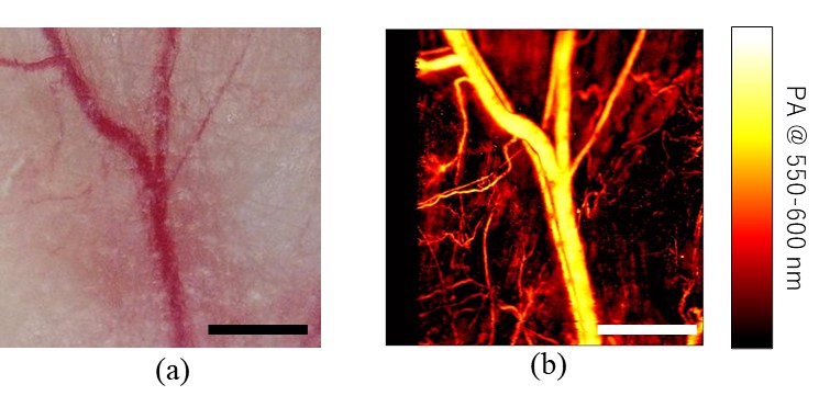 Blood vessels in mouse ear imaged by a photoacoustic microscopy (Hirasawa et al, J. Biomed. Opt., 29(S1), S11527, 2024)